 According to the Skin Cancer Foundation, skin cancer is the most common type of cancer in the United States. More than 9,500 people are diagnosed with skin cancer every day. More than two people die of the disease every hour.
One highly effective treatment for skin cancer lesions, particularly basal cell carcinomas and squamous cell carcinomas, is Mohs micrographic surgery. This procedure, named after its inventor Dr. Frederic Mohs, has a high success rate in treating basal cell carcinoma and squamous cell carcinoma, the two most common types of skin cancer.
At Peach Dermatology, we offer Mohs Micrographic Surgery as one of our treatment options for skin cancer. Let's take a closer look at what this surgery entails and why it is often the preferred choice for skin cancer treatment.
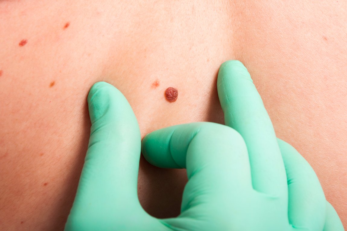
Common Signs of Skin Cancer
Skin cancer can present itself in many different ways, making it important to know the common signs and symptoms. These include:
- A new, changing, or unusual growth on the skin that does not go away within a few weeks. This could appear as a bump, spot, patch, sore, or mole.
- A sore that doesn't heal, or continually bleeds, scabs, or crusts.
- A mole that changes in size, shape, color, thickness, or texture over a short period of time.
- An existing mole that begins to itch, hurt, or become tender.
- Redness, swelling, tenderness or pain around a skin lesion.
The earlier you detect skin cancer, the easier it is to get rid of the cancer for good. If you notice any of these signs, it is important to have them checked by our dermatologist at Peach Dermatology as soon as possible.
Understanding Mohs Micrographic Surgery
Mohs micrographic surgery is a precise surgical technique used to remove skin cancer lesions while sparing as much healthy tissue as possible. It involves multiple stages of excision and examination of the tissue under a microscope to ensure complete removal of cancer cells. This process allows for the highest cure rate while minimizing the loss of healthy tissue.
During the procedure, a specially trained surgeon removes thin layers of skin one at a time, examining each layer under a microscope until no cancer cells are found in that layer. The entire surgery is typically completed in one day, and most patients can return home the same day.
What To Expect During the Procedure
Mohs micrographic surgery is typically performed under local anesthesia, meaning the patient is awake but their skin is numbed. The procedure itself may take several hours depending on the size and location of the lesion. Patients are often advised to bring reading materials or other forms of entertainment to keep them occupied during the waiting periods between tissue removal and examination.
Step 1: Examination and prep
The Mohs surgeon first inspects the biopsy site and marks the visible tissue with a pen. Depending on the cancer's location, you might sit up or lie down, covered with a surgical drape. If the cancer is on your face, your view might be blocked, but the surgeon will explain each step as it happens.
Next, the surgeon administers local anesthesia to numb the area thoroughly, ensuring you're awake but won't feel pain during the procedure.
Step 2: Tissue removal
Using a scalpel, the surgeon carefully removes the visible portion of the cancerous tissue along with a thin layer of surrounding tissue. Immediately after this step, the surgical area is temporarily bandaged. You'll then have time to rest as the removed tissue is sent for laboratory analysis.
Step 3: Lab analysis
In the lab, the surgeon divides the removed tissue into sections, using different colored dyes for identification, and creates a detailed map of the surgical area. A technician then freezes the tissue, slices it thinly like a layer cake, and places these slices onto microscope slides. Each slice is stained and covered for examination.
Step 4: Microscopic examination
The surgeon inspects the removed tissue for cancer cells, examining 100% of the surgical margin. If any are found, the surgeon marks their location on the map and removes another thin layer of tissue from that precise area. This process is repeated until no further cancer cells are found.
Step 5: Second layer removal
If any remaining cancer cells are detected, the surgeon administers additional anesthesia as required and precisely removes another skin layer from the identified area, as indicated by the map. This newly removed tissue is then sent to the lab for analysis, repeating the cycle until no more cancer cells are present.
Step 6: Reconstruction
Once your skin is free from cancer cells, your surgeon will discuss methods to repair or reconstruct the surgical wound. The type of reconstruction will depend primarily on the size and location of the lesion as well as your personal preferences. The wound may be closed with stitches, dressed with a bandage, skin graft, or flap. Regardless of the method used for closure, our priority is always to achieve optimal cosmetic and functional outcomes for our patients.
Step 7: Post-operative care
After the procedure, your surgeon will provide you with aftercare instructions to ensure proper healing. This may include keeping the surgical site clean and dry, avoiding strenuous activities or certain medications, and attending a follow-up appointment for monitoring and removal of any sutures or dressings. It is essential to follow these instructions carefully to avoid complications and optimize your recovery.
Why is Mohs Micrographic Surgery preferred?
There are several reasons why Mohs micrographic surgery is often the preferred treatment for skin cancer. Aside from a high cure rate, some of these reasons include:
- Precise removal of cancer cells: Mohs surgery removes only the affected tissue, leaving as much healthy skin intact as possible. This makes it an ideal option for skin cancers in delicate areas such as the face, ears, or nose.
- Minimizes scarring: By removing only the affected tissue and preserving surrounding healthy tissue, Mohs surgery results in minimal scarring compared to other treatment options.
- Cost-efficacy: While Mohs surgery may initially seem more expensive than other treatments, it is often a cost-effective option in the long run. This is because it has a higher success rate, reducing the chances of needing additional treatments in the future.
Choose the Best Skin Cancer Treatment!
Mohs micrographic surgery is a highly effective and precise treatment option for skin cancer. Its multi-stage process allows for the highest cure rate while minimizing the loss of healthy tissue. With minimal scarring, cost-effectiveness, and the ability to target delicate areas, it is no wonder why this procedure is often preferred by dermatologists and patients alike.
Peach Dermatology is proud to offer Mohs surgery as part of our comprehensive skin cancer treatment options.
Schedule a consultation with Dr. Shaikh to learn more about this procedure and determine if it is the right choice for you. You can also give us a call at 770-676-2200 or visit our office at 335 Peachtree Industrial Blvd, Suite 2210, Suwanee, GA 30024.
